- Planmeca Promax Service Manual
- Planmeca Promax 3d Mid User Manual Software
- Planmeca Promax 3d Mid Manual
- Planmeca Promax 3d Installation Manual
- Planmeca Promax 3d Mid Technical Manual
- Planmeca Promax 3d Mid User Manual 2016
- X-ray units with 3D sensor and SmartPan license: X-ray units with Dimax sensor: 22 Planmeca ProMax 3D Mid User’s Manual (2D) Page 27 The marking on the jaw icon demonstrates the image layer. Select the required jaw size by tapping the corresponding icon.
- Manufacturer’s Recommendations for Alternate Dental CBCT QA Program. Planmeca: ProMax 3D Max, 3D, 3Ds, and 3D Mid: Table 6. Medical Physicist’s Computed Tomography QC Survey. Item Required Test or Procedure Frequency. Planmeca Substitute Test or Procedure Standard. 1 Scan Increment Accuracy.
Planmeca ProMax® Quick guide – Capturing 2D image Preparation Planmeca Romexis® software 1 Select Panoramic exposure. Planmeca ProMax® X-ray unit 2 Select 2D Dental. 3 Select Panoramic. 4 Select program type. 3 4 2 5 Select patient size. 6 Apply segmentation if needed. 7 Gwod.r oarf 5 6 7 General instructions during exposure The patient.
DESCRIPTION
Proline XC X-rays are designed for full-view patient positioning...Proline XC X-rays are designed for full-view patient positioning using a three laser positioning light system and a Graphic User Interface with easy-to-follow, color-coded menu options. The XC's focal trough adjusts to the patient's jaw shape and size providing excellent image quality. In addition, the Proline XC can be upgraded with future software features by simply replacing a software chip! With over 10+ programs from which to choose, the XC is definitely an economical solution for excellent dental imaging.
FORUMSView All (8)
Ask a New Question0Replies10 months ago | 10 months agotroubleshooting message on computer after taking pano I took a pano today and the pano worked but did not come up on the computer and the message says no license on fileReply |
| -Del Mar View Dental Care 2 years ago | 2 years agoit wont rotate properly, off center at start up. It wont finish it wont rotate properly, off center at start up. It wont finishReply |
0Replies3 years ago | 3 years agoPlanmeca ProMax We have a Promax that the images produces a white line at mid-line during exposure. We are exporing possible solutions to the white out at mid-line. Reply |
DOCUMENTS / MANUALSView All
VIDEOS
FEATURES
- Open and easy patient access
- Comfortable and stable patient supports
- Side entry and open view for practical and precise patient positioning
- Triple laser beam system for accurate alignment of reference anatomical landmarks
- TheGraphicUserInterface(GUI)for intuitive selection of exposure program and parameters
- With state-of-the-art direct digital imaging the image is available for diagnosis immediately after exposure
Additional Specifications
Generator: Constant potential, microprocessor controlled, operating frequency 80 kHz
X-ray tube: D-052SB
Focal spot size: 0.5 x 0.5 mm
Total filtration: 2.5 mm Al
Anode voltage: 60 - 80 kV
Anode current: 4 - 12 mA DC
Exposure time:
Pan-2.5 - 18 s
Ceph- 0.2 - 5 s
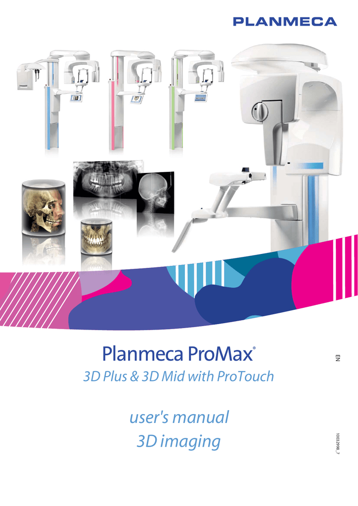
Planmeca ProMax® 3D Mid is a genuine all-in-one CBCT (Cone Beam Computed Tomography) unit including 3D imaging, 3D photo, digital 2D panoramics and cephalometry, all in the same unit. Planmeca ProMax® 3D Mid complies with a multitude of diagnostic requirements: those of implantology, endodontics, periodontics, orthodontics, dental and maxillofacial surgery, and TMJ analysis. It is also an excellent tool for diagnosing ear, maxillary sinus, and… Read more
Promotions
Literature
Description
Planmeca ProMax® 3D Mid is a genuine all-in-one CBCT (Cone Beam Computed Tomography) unit including 3D imaging, 3D photo, digital 2D panoramics and cephalometry, all in the same unit.
Planmeca ProMax® 3D Mid complies with a multitude of diagnostic requirements: those of implantology, endodontics, periodontics, orthodontics, dental and maxillofacial surgery, and TMJ analysis. It is also an excellent tool for diagnosing ear, maxillary sinus, and respiratory tract diseases.
Planmeca ProMax 3D Mid provides volumes sizes for every clinical application with the possibility to adjust the volume position according to acquired scout images.
Adjustable
volume sizes and resolution modes
- Volume sizes from Ø34×42 mm to Ø200×170 mm
Unique 3D combination – an industry first
We’re the first company to combine three different types of 3D data with one X-ray unit. The Planmeca ProMax® 3D family brings together a Cone Beam Computed Tomography (CBCT) image, 3D face photo and 3D model scan into one 3D image – using the same advanced software. This 3D combination creates a virtual patient in 3D, helping you with all your clinical needs.

Pioneering Planmeca Ultra Low Dose™ protocol. An even lower patient dose than in panoramic imaging
Planmeca ProMax® 3D units offer a unique Planmeca Ultra Low Dose™ imaging protocol, enabling CBCT imaging with an even lower effective patient dose than standard 2D panoramic imaging. This pioneering imaging protocol is based on intelligent 3D algorithms developed by Planmeca and offers a vast amount of detailed anatomical information at a very low patient dose.
Planmeca Promax Service Manual
Planmeca ProMax® 3D family
Key features
Advanced technology:
- Ideal resolutions and patient dose levels that always comply with the ALARA (As Low As Reasonably Achievable) principle
- Optimal volume size and location for every clinical need
- Special imaging protocols for dental and ENT applications
Effortless use:
- Effortless patient positioning and unmatched comfort
- True all-in-one X-ray units not only for 3D imaging, but 2D panoramic and cephalometric imaging as well
- Easy to use for a smooth workflow
- Planmeca Romexis® software
- Mac OS and Windows support
Ease of operation
Planmeca Promax 3d Mid User Manual Software
Our Planmeca ProMax® 3D units are known across the world for incredible ease of use and exceptional patient comfort.
A relaxed patient means a smooth imaging workflow and the best quality images.
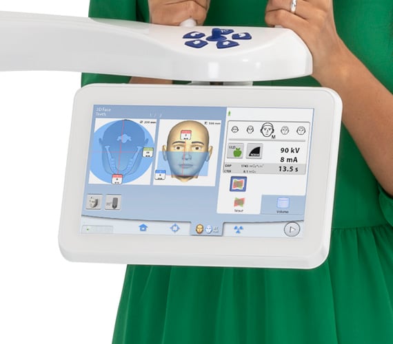
User-friendly control panel
- Clear and straightforward interface guides users smoothly through their work
- Pre-programmed sites and exposure values for different image types and targets saves time and allows full focus on patients
Open patient positioning
Planmeca Promax 3d Mid Manual
- Effortless positioning with open-face architecture
- Unrestricted view of patients
- Calm experience for patients – without claustrophobic feelings
- Fine adjustment using positioning lasers and a joystick
- Correct positioning verified with a scout image
- Easy wheelchair accommodation with side-entry access
Specifications
Technical data

- Anode voltage 54–90 kV
- Anode current 1–12 mA
- Focal spot 0.5 mm, fixed anode
- Image detector Flat panel
- Image acquisition 200/360 degree rotation
- Scan time 2–33 s
- Reconstruction time 2–55 s
Dimensions
3D Plus or 3D Mid
A 1315–2095 mm (51.8–82.5 in.)
B 1610–2390 mm (63.4–94.1 in.)
C 1130 mm (44.6 in.)
D 930 mm (36.6 in.)
E 247 mm (9.7 in.)
F 810 mm (32 in.)
G 1366 mm (53.8 in.)
H 756 mm (29.8 in.)
J Ø1010 mm (39.8 in.)
Physical space requirements
3D Plus or 3D Mid
Width 118 cm (47 in.)
Depth 137 cm (54 in.)
Height** 161–239 cm (64–94 in.)
Weight 131 kg (lbs 289)
3D Plus or 3D Mid with cephalostat
Width 206 cm (82 in.)
Depth 137 cm (54 in.)
Height** 161–239 cm (64–94 in.)
Weight 146 kg (lbs 322)
Minimum operational space requirements
Planmeca Promax 3d Installation Manual
3D Plus or 3D Mid
Width 158 cm (63 in.)
Depth 175 cm (69 in.)
Height** 239 cm (94 in.)
Planmeca Promax 3d Mid Technical Manual
3D Plus or 3D Mid with cephalostat
Planmeca Promax 3d Mid User Manual 2016
Width 225 cm (89 in.)
Depth 175 cm (69 in.)
Height** 239 cm (94 in.)
**The maximum height of the unit can be adjusted for offices with limited ceiling space.